
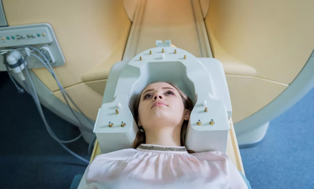
An MRI scan shows where a brain tumour is and how big it is. But does it say how dangerous the cancer is and whether treatment will work? Giacomo Annio tests tumours with vibrations: a device the size of a phone, placed against the head, shows whether tissue is stiff or soft. The result is surprising, because the most aggressive glioblastomas are softer than their surroundings — and as this is a story about looking deeper, at the end you can check whether you judge by appearances yourself.
An MRI scan can show how big a brain tumour is and exactly where it sits. It is less helpful with what a doctor often needs most: the biological properties of the tumour, which decide whether treatment will work. The image tells you where and how big, but says little about what kind of tumour it is.
Giacomo Annio, a fellow of the Marie Skłodowska-Curie Actions programme, set out to fill that gap. In the GLIOBID project he studied glioblastomas, the most aggressive group of brain tumours. His tool is magnetic resonance elastography. The three-year project was coordinated by Oslo University Hospital, which is where the clinical data came from. Preclinical research was carried out at Stanford University.
The idea is simple: instead of only looking at the tumour, find out what it is like to the touch. “Elastography measures how stiff or soft [tumour] tissue is,” explains Annio. As he adds, these very properties offer clues to how the tumour behaves, whether it resists therapy and whether it infiltrates surrounding tissue.
According to the researcher, such a method lets doctors look at a tumour from a new perspective and analyse its physical properties in a way that an ordinary image does not allow.
The equipment itself is nothing like a huge machine. The elastography device is the size of a mobile phone. The doctor places it against the patient’s head, and the waves it sends into the brain with its vibrations are visualised by an MRI scanner.
The design is being developed by the American company Quality Electrodynamics, and Germany’s Siemens commercialises it on its scanners. The solution is beginning to appear in the first hospitals. That does not mean everything is ready, though. “Adapting it for brain tumours is a particular challenge,” Annio admits.
GLIOBID did not come out of nowhere. It builds on two earlier projects: FORCE, which developed elastography, and ImPRESS, which applied it to brain tumours. Annio was part of both teams.
The researcher spent the first two years of his fellowship in the Molecular Imaging Program of the Department of Radiology at Stanford University. There he built his own elastography device using 3D printing and customised electronics, the first of its kind at Stanford. It was installed on an MRI scanner and used to study brain tumours in mice.
An important outcome of the work at Stanford is a patent. The team patented a theranostic agent, a molecule that combines therapy and diagnosis and can be seen on MRI. “This molecule releases a component that changes the mechanics of the tumour, opening up the blood vessels specifically at the tumour site,” Annio describes. Wider vessels mean that more treatment can reach the tumour.
Elastography also made it possible to follow what was happening in the tissue. “Using elastography, we could see how the mechanical properties of the tumour impact on the delivery of therapy,” says the researcher. In his view this is a first: “No one had done this before.”
The results so far concern animals, but they show the direction well. In short:
The effectiveness of the therapy has so far been shown only in mice. This is still the preclinical stage.
The second part of the work relied on human data. A clinical study at Oslo University Hospital involved 200 patients with glioblastoma. It turned out that glioblastomas break the rule. “While most cancers are stiffer than surrounding tissue, brain glioblastomas are softer,” says Annio.
The researcher believes that in judging how aggressive a tumour is, its viscosity, which he calls ‘squeezability’, may matter more than its stiffness. In other words: it counts not only whether the tumour is hard, but also how well it can be squeezed.
Elastography also sheds light on the blood vessels inside the tumour and lets doctors follow the pressure building up inside the brain. A growing tumour squeezes its vessels, which in this crowded space cannot properly deliver blood, oxygen, nutrients or treatment.
The method has also been extended to meningioma, one of the more common brain tumours. Kyrre Eeg Emblem, head of the Department of Physics and Computational Radiology at Oslo University Hospital and project coordinator, points to the practical side of this work. As he explains, “the assessment of physical forces allows the neurosurgeon to plan surgery” by reviewing whether the tumour will be easy or difficult to remove.
An ordinary MRI scan shows the shape and location of a brain tumour, while elastography adds information about its physical properties. In the GLIOBID project, glioblastomas turned out to be softer than the surrounding tissue, and what may be key is their squeezability. In mice, combining elastography with a patented theranostic molecule shrank the lesions and increased survival. The treatment results so far concern animals, but the direction is clear: therapy matched to what a tumour is like to the touch.
5 questions · one minute


 EN

 CS
 DE
 ES
 FR
 HU
 IT
 PL
 PT
 РУ
 SK
 TR
 УК
 AR
 中文