
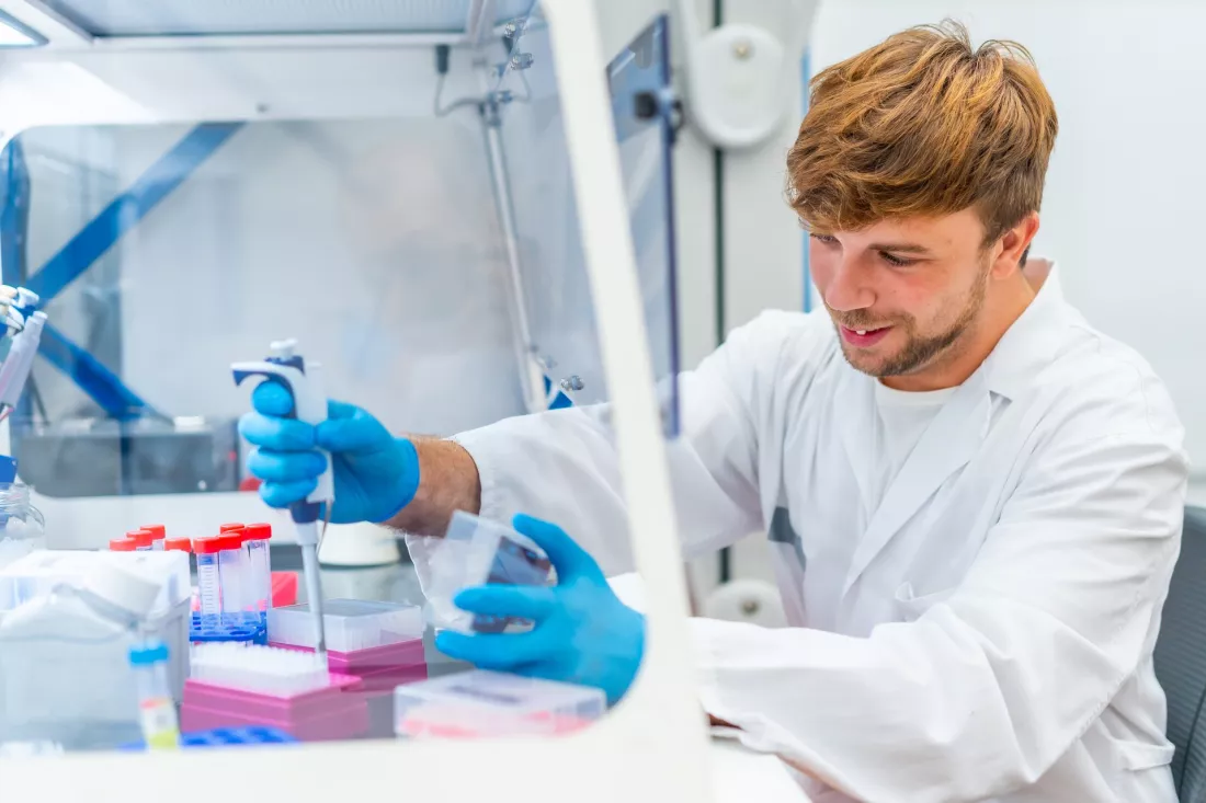
Coffee waste and peanut shells normally end their life in a bin. In a European project coordinated from Lyon they became the raw material for the carbon cores of nanoparticles smaller than twenty nanometres — particles that show a tumour on an image and attack it at the same time. In animal studies the team reports up to 97% inhibition of tumour growth. It is a story about materials chemistry, biology and imaging meeting inside one object — and at the end you can check how you look at things everyone else calls waste.
In most hospitals, finding a tumour and treating it belong to two different worlds. One machine produces the image, another team plans the therapy, and the patient travels between them. As the report on the UNAT project puts it, "Cancer diagnosis and treatment often rely on separate technologies, meaning that clinicians may need different procedures to locate tumours, assess their characteristics and deliver treatment."
That split costs time, and every additional procedure is another load on someone who is already ill. "Developing safer tools that can combine these functions could help make cancer care more precise while reducing the burden on patients." The project, funded by the Marie Skłodowska-Curie Actions, set out to build exactly such a tool.
The surprising part is where the building material comes from. The heart of every particle is carbon, and carbon does not have to arrive from a chemical catalogue. "Carbon cores can be produced from biomass and biowaste, including coffee waste and peanut shells, providing an alternative source for the production of nanomaterials."
So the shopping list of a laboratory working on next-generation oncology includes:
Nothing on that list is exotic, and everything on it is normally thrown away.
The nanohybrids built in the project stay below 20 nanometres. At that scale size stops being a technical detail and starts deciding everything: how a particle moves through the body, how deep it reaches, and whether the body can later get rid of it. Most of what follows in this story is a consequence of that one number.
The same object carries more than one way of being seen. Multispectral fluorescence makes it possible to watch what happens at the level of cells, while high-contrast magnetic resonance imaging reaches deep into tissue, where light does not go. Instead of one contrast agent for the scanner and a separate dye for the microscope, there is a single platform that answers both questions.
And it does not stop at looking. The same particle carries the therapeutic part of the job, which is why the researchers talk about diagnostics and treatment merging rather than following one another.
Every nanomaterial that enters a body raises the same question: where does it end up in a year. Here the answer comes from the ultrasmall size and the negative charge of the surface. The particles are cleared quickly by the kidneys and through the hepatobiliary route, so they do not accumulate in organs, and the risk of long-term toxicity drops.
The tests in mice produced the figures that carry this project. A nitrogen-rich version of the nanohybrids reached 93% inhibition of tumour growth, together with fewer lung metastases. A version containing gadolinium worked as a radiosensitiser, making the tumour more vulnerable to radiation, and reached 97% inhibition of tumour growth.
"Carbon-based nanohybrids developed in the UNAT project represent a highly promising next-generation medical tool because they seamlessly combine advanced diagnostics and targeted therapy into a single, ultrasmall platform," says project coordinator Vladimir Lysenko.
UNAT is a good argument against choosing a field too early. The carbon core is materials chemistry. The behaviour of the particle in an organism is biology and pharmacology. The signal it sends back is imaging diagnostics and physics. None of these subjects alone produces the result, and all of them meet inside one object smaller than a virus.
There is also a second reason, less technical. Medicine measured on population averages tells a patient what usually happens. A tool that images and treats at once opens a different logic: "Rather than relying solely on population-level responses, such an approach could help identify patients most likely to benefit from a particular treatment."
UNAT is coordinated by Université Lyon 1 Claude Bernard in France, runs from 1 April 2021 to 31 March 2026 under grant agreement 101008159, and has a budget of EUR 832 600. Before anything reaches a hospital, the team still has manufacturing, toxicology and formal trials ahead.
A European project turned coffee waste and peanut shells into carbon cores of particles under 20 nanometres that image a tumour and treat it at the same time, with 93% and 97% inhibition of tumour growth in animal studies and fast clearance from the body. The route from a bin to a medical tool runs through three different degree programmes at once, which is the most useful thing a student can take from this story.
5 questions · one minute · nothing is saved


 EN
EN 
 CS
CS DE
DE ES
ES FR
FR HU
HU IT
IT PL
PL PT
PT РУ
РУ SK
SK TR
TR УК
УК AR
AR 中文
中文