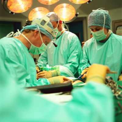
A ‘smart’ knife to fight cancer, crime and contamination

Surgical operation
Cancer is one of the most challenging medical issues we face. In the United Kingdom alone, there are 300 000 new cases every year — leading to almost 2 million surgical operations annually. Thanks to ERC funding, Dr Zoltán Takáts of Imperial College London has developed a ‘smart’ surgical knife that can ‘smell’ the tissues it is cutting through — with the potential to revolutionise cancer treatment, as well as food and drug analysis, and research into the human ‘microbiome’.
The instrument, which the researchers call the
‘iKnife’, uses a mass spectrometer to analyse
the ‘smoke’ produced by a scalpel that cuts
using high-frequency electric current. From
this, surgeons receive real-time feedback on
whether the tissue they are cutting is cancerous or healthy.
Dr Takáts’ project, DESI-JEDI IMAGING 1 ,
has already reported a successful trial of
the technology in the journal Science of
Translational Medicine, which attracted a
great deal of press attention. During the
study, the project built up a database of signatures for different cancers — including
brain, breast, lung and colonic — by taking
tissue samples from more than 300 patients.
The iKnife was then used in 80 surgical
operations, and in every case its real-time
results matched the traditional tissue analysis
carried out after the operation.
‘We didn’t expect to get this far in the project
when we made the original proposal in 2007,’
says Dr Takáts. ‘But in the six weeks since our
paper was published we’ve gone on to complete our clinical experiments and have shown
the technology is ready for application — so
we are now close to beginning official clinical
trials with a view to regulatory approval.’
A beautiful breakthrough
Mass-spectrometers measure the mass-to-
charge ratio of ionised (charged) particles
by passing them through electric or magnetic fields. Scientists use them to analyse
the chemical composition and structure of
samples.
The big breakthrough for Dr Takáts came
when he realised that new surgical techniques — such as ultrasound-, laser or electrosurgery — produced charged particles of
tissue which were ideally suited as input to
a mass spectrometer. His first simple experiment — using pork liver and off-the-shelf
surgical instruments — worked beautifully,
with results much better than expected.
‘For this fantastic tool, we immediately
started thinking about applications — we
looked for problems where an imaging mass
spectrometer could help transform the
landscape.’
Currently, a surgeon would take a tissue sample and have it analysed (called a ‘biopsy’) by
a histopathology laboratory — with at least
a 40-minute wait — before knowing whether
to continue the operation. But by using an
air pump to ‘suck’ particles from the site of
the surgery to the mass spectrometer — the
iKnife can supply instant feedback to the sur-
geon so that he or she can avoid cutting away
more tissue than is necessary to remove all
tumorous or inflamed cells. Leaving more
of the remaining tissue intact will improve
medical outcomes and patients’ quality of
life.
From science to surgery
‘We are now moving to official clinical trials,’
says Dr Takáts. ‘We expect to see the start of
trials in brain surgery early next year.’
This has proved a challenge, since the design
of all three of the instrument’s components — the electro-scalpel, the database for
identification and diagnosis, and the mass
spectrometer tailored for the operating theatre — must be finalised before clinical trials
begin because it cannot be changed after
approval.
‘The ERC’s Proof-of-Concept (PoC) Grant was
of key importance to this,’ says Dr Takáts.
‘The Starting Grant gave us a huge opportunity to set up the research group and do the
science, but we really needed the PoC funding to look into regulatory issues, intellectual
property management and starting up a company to bring the instrument to market.’
The iKnife has been developed for electro-
surgery, as this is preferred by cancer specialists, but the technique is also applicable to
hydro- and laser surgery. New applications for
the tool could also include analysis of mucous
membranes and the respiratory, urinogenital
or gastrointestinal systems.
Its usefulness for tissue and substance analysis
has led the team to have talks with food
industries and crime-fighting agencies. It could
also be a powerful new tool for microbiologists,
accelerating the creation of databases of the
microbial population inhabiting our bodies.
‘The millions of bacteria that live in — and on
— us may be linked to cancers and illnesses
like diabetes,’ says Dr Takáts. ‘Identifying
them may help in diagnosis and treatments.
We now have a unique tool to look into the
fine detail of those interactions — leading to
new approaches and therapies.’
The project was coordinated by Imperial
College, London, in the United Kingdom.
published: 2014-08-31